
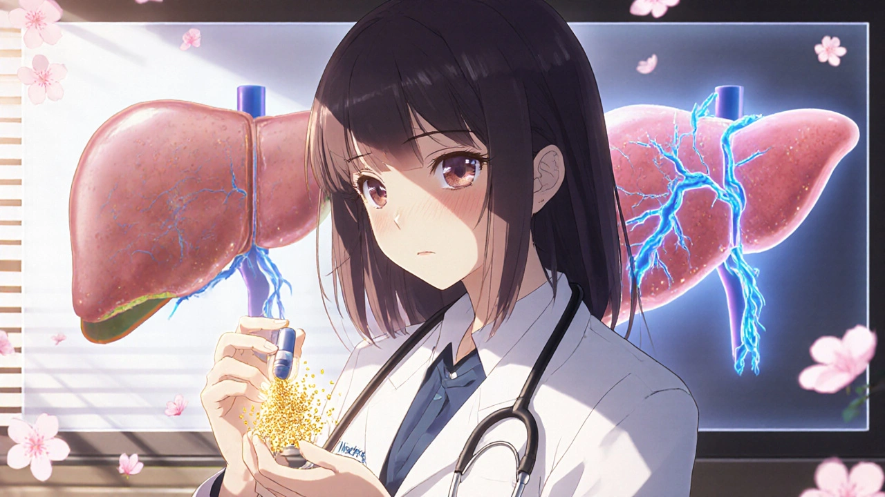
What Is Portal Vein Thrombosis?
Portal vein thrombosis (PVT) happens when a blood clot blocks the portal vein - the main vessel that carries blood from the intestines to the liver. It’s not rare, especially in people with liver disease, cancer, or inherited clotting disorders. The clot can be partial or complete, and it can form suddenly (acute) or develop over time (chronic). Acute PVT shows up fast, often with abdominal pain or bloating. Chronic PVT builds up slowly, sometimes without symptoms until complications like portal hypertension or varices appear.
First described in 1868 by Rudolf Virchow, PVT follows his classic triad: blood that clots too easily, damaged blood vessel lining, and slow blood flow. Today, we know that cirrhosis is the top cause - but it’s not the only one. Other triggers include recent abdominal surgery, pancreatitis, infections like appendicitis, and genetic clotting conditions like Factor V Leiden. About 25-30% of non-cirrhotic patients have an underlying clotting disorder. Without treatment, the clot can spread into the intestines, cutting off blood supply and leading to life-threatening ischemia.
How Is Portal Vein Thrombosis Diagnosed?
Ultrasound is the first test doctors reach for. It’s fast, cheap, and picks up PVT in 89-94% of cases. A Doppler ultrasound shows whether blood is flowing normally through the portal vein. If the vein looks blocked, the clot’s size and location are measured: less than 50% blockage is minimal, 50-99% is partial, and 100% is complete. When the original vein disappears and is replaced by a web of tiny vessels - called cavernous transformation - it’s a sign of chronic PVT.
If ultrasound is unclear, a CT scan or MRI with contrast gives a clearer picture. These show exactly how far the clot extends and whether it’s reached branches inside the liver or into the spleen or intestines. Blood tests alone won’t confirm PVT, but they help assess liver function. Child-Pugh and MELD scores are used to judge how bad the liver damage is. A high score means higher bleeding risk, which changes how aggressively you treat the clot.
Doctors also check for varices - swollen veins in the esophagus or stomach - using endoscopy. These are common in cirrhotic patients with PVT and can bleed suddenly if not treated before starting anticoagulants. A thrombophilia workup (testing for Factor V Leiden, prothrombin mutation, protein C/S deficiency) is recommended for non-cirrhotic patients, especially if they’re young or have no obvious cause.
Why Anticoagulation Is the Standard of Care
For years, doctors were hesitant to use blood thinners in patients with liver disease, fearing bleeding. But evidence has flipped that thinking. Today, major guidelines from AASLD and EASL agree: anticoagulation is the standard for most acute PVT cases, even in cirrhosis - if managed carefully.
The goal isn’t just to dissolve the clot. It’s to stop it from growing, prevent intestinal damage, and let the vein reopen. Studies show that patients treated within six weeks have a 65-75% chance of full or partial recanalization. Those who wait? Only 16-35% see improvement. Untreated PVT cuts 5-year survival to under 50%. With anticoagulation, it jumps to 85%.
For non-cirrhotic patients, DOACs (direct oral anticoagulants) like rivaroxaban and apixaban now lead the pack. In one 2020 study, 65-75% of patients on DOACs had complete clot resolution. That’s far better than warfarin’s 40-50%. DOACs are easier to use - no regular blood tests, fewer food interactions. But they’re not for everyone. If the liver is severely damaged (Child-Pugh C), DOACs are still avoided.
Choosing the Right Anticoagulant
Not all blood thinners are equal in PVT. The choice depends on liver function, bleeding risk, and whether the patient has cancer or a clotting disorder.
- LMWH (low molecular weight heparin) - Like enoxaparin - is often used first, especially in cirrhosis. It’s given as a daily or twice-daily shot. Dose is based on weight: 1 mg/kg twice daily or 1.5 mg/kg once daily. Target anti-Xa levels: 0.5-1.0 IU/mL. It’s stable in liver disease and has fewer bleeding risks than warfarin in Child-Pugh A/B patients.
- Vitamin K antagonists (VKAs) - Warfarin is the main one. Requires frequent INR checks. Target: 2.0-3.0. Works well, but harder to control. In cirrhotic patients, recanalization rates are lower (30-40%) compared to LMWH (55-65%).
- DOACs - Rivaroxaban (20 mg daily), apixaban (5 mg twice daily), dabigatran. No routine monitoring needed. Best for non-cirrhotic patients. Recanalization rates: 65-75%. FDA warns against DOACs in Child-Pugh C. But new data from the 2024 AASLD update now supports DOACs in Child-Pugh B7 patients, based on the CAVES trial showing non-inferior results to LMWH.
For patients with active cancer, LMWH remains first-line. For those with inherited clotting disorders, lifelong anticoagulation is usually needed. Treatment duration? At least six months for provoked cases (like after surgery). Lifelong for unprovoked or genetic causes.
When Anticoagulation Is Too Risky
Anticoagulation saves lives - but it can also cause serious bleeding. In cirrhotic patients, major bleeding happens in 5-12% of cases, mostly from ruptured varices. That’s why screening is non-negotiable.
Anticoagulation is avoided if:
- The patient had a recent variceal bleed (within 30 days)
- They have uncontrolled ascites or high portal pressure
- They have Child-Pugh class C cirrhosis
- Platelets are below 50,000/μL (though some centers safely treat with transfusions to raise counts above 30,000)
Experts like Dr. Dominique Valla recommend endoscopic band ligation before starting anticoagulation in cirrhotic patients. UCLA’s 2022 study showed this cut major bleeding from 15% to just 4%.
Also, don’t start anticoagulation blindly in asymptomatic PVT without cancer or symptoms. Dr. Paul Angulo’s 2022 editorial reminds us: not every clot needs thinning. The risk might outweigh the benefit.
What If Anticoagulation Fails?
Some clots don’t respond to blood thinners. Or they cause severe complications like intestinal ischemia. When that happens, doctors turn to more invasive options.
- TIPS (Transjugular Intrahepatic Portosystemic Shunt) - A metal tube placed between the portal and hepatic veins to reroute blood. Success rate: 70-80%. But 15-25% of patients develop hepatic encephalopathy. Used after anticoagulation fails.
- Surgical shunts - Open surgery to create a bypass. Rare now due to TIPS and higher complication rates.
- Percutaneous thrombectomy - A catheter is threaded in to physically remove the clot. Immediate recanalization in 60-75% of cases. Only available at specialized centers.
These are last-resort options. They’re risky and expensive. But for patients with mesenteric ischemia or transplant candidates with large clots, they can be lifesaving. At UCSF, anticoagulation reduced transplant exclusion due to PVT from 22% to just 8%.

Real-World Outcomes and Challenges
At Mayo Clinic, 78% of patients treated within 30 days saw clot improvement. Only 42% did if treatment was delayed. At Cleveland Clinic, 18% of Child-Pugh B patients stopped anticoagulation because of bleeding fears. That’s a big problem - fear is costing lives.
One key lesson from Johns Hopkins: multidisciplinary teams work best. Hepatologists, radiologists, transplant surgeons, and nurses coordinating care reduced complications by 35%. That’s not magic - it’s structure.
Another challenge? Many doctors aren’t trained in PVT management. Only 35% of general gastroenterologists feel confident handling anticoagulation in PVT, according to a 2023 ABIM survey. Community hospitals often lag behind academic centers. Only 45% of community hospitals have formal PVT protocols, versus 82% of transplant centers.
And timing matters. Massachusetts General found 22% of patients who presented with intestinal ischemia died - versus 5% in those treated early. The window to act is narrow.
What’s Next for PVT Treatment?
The field is changing fast. DOACs are replacing warfarin in non-cirrhotic cases. By 2025, 75% of those cases are expected to use DOACs. New data supports their use even in some compensated cirrhotic patients.
The 2023 FDA approval of andexanet alfa - a reversal agent for rivaroxaban and apixaban - is a game-changer. Now, if a patient bleeds, there’s a targeted antidote. That gives doctors more confidence to treat.
Future therapies are coming. Abelacimab, a new anticoagulant in phase 2 trials, shows promise for patients who can’t tolerate current drugs. Targeted thrombolysis - dissolving clots with drugs delivered directly to the clot - is gaining traction and could reach 15% market use by 2027.
Genetic testing is also becoming part of routine care. Patients with Factor V Leiden or prothrombin mutation have 80% higher recanalization rates with long-term anticoagulation. That means treatment can be personalized - not one-size-fits-all.
Getting Started: A Practical Checklist
If you suspect PVT, here’s what to do:
- Get a Doppler ultrasound - confirm the clot.
- Check liver function - calculate Child-Pugh and MELD scores.
- Screen for varices with endoscopy - treat them before starting anticoagulants.
- Test for thrombophilia - especially if no clear cause.
- Choose anticoagulant based on liver status: DOACs for non-cirrhotic, LMWH for cirrhotic.
- Start treatment ASAP - within days, not weeks.
- Monitor for bleeding - especially in cirrhosis.
- Reassess at 3-6 months with imaging to check for recanalization.
Don’t wait. The earlier you treat, the better the outcome. And don’t be afraid to consult a liver specialist. PVT is complex - but with the right approach, it’s manageable.
Can portal vein thrombosis be cured?
Yes, in many cases. With early anticoagulation, 65-75% of patients achieve partial or complete recanalization of the portal vein. Recanalization means the clot dissolves or is absorbed, and blood flow returns. This is most likely when treatment starts within six weeks. Chronic PVT is harder to reverse, but anticoagulation still prevents worsening and reduces complications like variceal bleeding.
Is anticoagulation safe for people with cirrhosis?
It can be - if done carefully. For Child-Pugh A and B cirrhosis, anticoagulation is now recommended, especially for acute PVT. LMWH is preferred over warfarin because it’s more predictable. DOACs are approved for Child-Pugh B7 patients as of early 2024. But they’re still avoided in Child-Pugh C. The key is screening for varices first and avoiding anticoagulation if there’s recent bleeding or uncontrolled ascites.
Do I need lifelong anticoagulation for portal vein thrombosis?
Not always. If the clot was triggered by something temporary - like recent surgery or infection - six months of treatment is usually enough. But if you have an inherited clotting disorder (like Factor V Leiden), active cancer, or unprovoked PVT with no clear cause, lifelong anticoagulation is recommended. Your doctor will decide based on your risk of recurrence.
Can portal vein thrombosis lead to liver failure?
Not directly, but it can make liver disease worse. PVT increases pressure in the portal system, which can worsen ascites, varices, and liver function. In transplant candidates, large clots can make them ineligible for transplant. Anticoagulation improves post-transplant survival - studies show 85% one-year survival in treated patients versus 65% in untreated ones. So treating PVT helps protect the liver, even if it doesn’t reverse cirrhosis.
What are the signs that PVT is getting worse?
Watch for sudden abdominal pain, fever, nausea, vomiting, or bloody stools - these could mean intestinal ischemia. Worsening ascites, new variceal bleeding, or confusion (hepatic encephalopathy) suggest rising portal pressure. If you’re on anticoagulation and notice unusual bruising, dark stools, or dizziness, that could signal bleeding. Any new or worsening symptoms need immediate medical attention.

Comments (10)
Frances Melendez
This is why people die in this country-doctors still don’t take PVT seriously until it’s too late. I’ve seen it firsthand. My aunt had chronic PVT for years and was told to ‘wait and see.’ She ended up with a bowel infarction. Anticoagulation isn’t optional. It’s basic. Stop playing doctor and start saving lives.
Jonah Thunderbolt
OMG 😱 this post is literally a MASTERCLASS 🏆. I mean, who even wrote this? A hepatologist? A wizard? DOACs in Child-Pugh B7?? 🤯 The CAVES trial?? I’m crying. This is the kind of content that makes me believe medicine still has hope. 10/10 would recommend to my ENTIRE family. Also, if you’re not using rivaroxaban for non-cirrhotic PVT, you’re basically using a flip phone in 2024. 📱➡️📱💥
Rebecca Price
I appreciate how thorough this is. Really. But I also wonder-how many patients never even get to this level of care? I work with underserved communities, and the idea of ‘get a Doppler ultrasound within days’ sounds like a fantasy when your nearest imaging center is 90 miles away. We need systemic change, not just clinical guidelines. This info is gold-but it’s useless if it doesn’t reach the people who need it most.
marie HUREL
I’ve been following PVT research since my dad had it after abdominal surgery. The shift from warfarin to DOACs was huge for us-no more monthly INRs, no more broccoli restrictions. I still remember the relief when his follow-up scan showed partial recanalization. It’s not a cure, but it’s a second chance. Thank you for sharing the practical checklist. I’m printing it out.
Gayle Jenkins
Let me cut through the noise: if you’re not screening for varices before anticoagulation, you’re not a doctor-you’re a liability. Endoscopic band ligation isn’t optional. It’s the gatekeeper. And yes, I’ve seen patients bleed out because someone was ‘afraid’ to treat. Fear kills more than clots. Stop being timid. Start being competent. This isn’t theory. It’s survival.
Cecily Bogsprocket
I keep thinking about how much of medicine is still shaped by fear. Fear of bleeding. Fear of doing too much. Fear of being wrong. But what’s the cost of inaction? A 50% five-year survival rate? That’s not cautious-it’s cruel. We treat cancer with everything we’ve got. Why not this? The data doesn’t lie. The real question is: why are we still hesitating?
Rhiana Grob
The mention of andexanet alfa as a game-changer is spot on. For years, we’ve been afraid to anticoagulate cirrhotic patients because there was no antidote. Now, we have one. That changes the risk-benefit calculus entirely. I predict we’ll see a 40% increase in anticoagulation initiation in the next two years. This is the dawn of a new era.
Savakrit Singh
It is imperative to note that the utilization of direct oral anticoagulants (DOACs) in non-cirrhotic patients must be contextualized within the framework of renal function, as creatinine clearance thresholds are not universally addressed in current guidelines. Furthermore, the CAVES trial’s exclusion criteria-particularly regarding albumin levels and platelet function-render its applicability to real-world populations questionable. One must exercise extreme caution before adopting DOACs as first-line in borderline Child-Pugh B patients without individualized pharmacokinetic assessment.
sharicka holloway
My cousin had PVT after pancreatitis. She was 32. No cirrhosis. No history. Just… a clot. They started her on Lovenox right away. Six months later, her vein was wide open. No one told her it could be reversed. No one. This post? It’s the thing I wish someone had handed me. Thank you.
Alex Hess
This is just another overhyped medical blog disguised as science. DOACs aren’t magic. They’re expensive. And no, not every patient needs anticoagulation. You’re pathologizing incidental findings. I’ve seen 3 asymptomatic PVTs in 10 years. All were stable. All were left alone. The real epidemic here? Over-treatment.